






- Home
- About ICCBS
-
Centers
- H.E.J. Research Institute of Chemistry
- Dr. Panjwani Center for Molecular Medicine & Drug Research
- Other Chemistry Centers
- Other Biological Sciences Related Centers
- Third World Center for Science and Technology
- Center for Bioequivalence Studies and Clinical Research (CBSCR)
- Industrial Analytical Center (IAC)
- Biotechnology Wing
- Prof. Dr. Wolfgang Voelter Laboratories Complex (NRL)
- Welcome to National Facility for Laboratory Animal Research and Care (NFLARC)
- L.E.J. Nanotechnology Center
- Technology Park & Technology Incubation Center
- Latif Ebrahim Jamal National Science Information Center
- Jamil-ur-Rahman Center for Genome Research
- National Institute of Virology (NIV)
- Sindh Forensic DNA and Serology Lab (SFDL)
- Academics
- Facilities
- Libraries
- Campus Life
- Newsletters
- Performance Report
- Alumni
- Conference
MICROSCOPE IMAGING FACILITY
MICROSCOPE IMAGING FACILITY

a:1:{s:7:"English";s:2533:"
";}
Robert Hooke and Anton van Leeuwenhoek devised the simple microscope to have better look at the cells never seen before. Microscopes and its associated techniques aided the human eye to observe the world of unseen microbes, tissue structures, and cellular complexities which led to great discoveries over the years. Microscopes are fundamental to study the complexity of cells and their metabolic processes. Since the discovery of compound microscope huge development have been made to improve the resolution of microscopic devices.
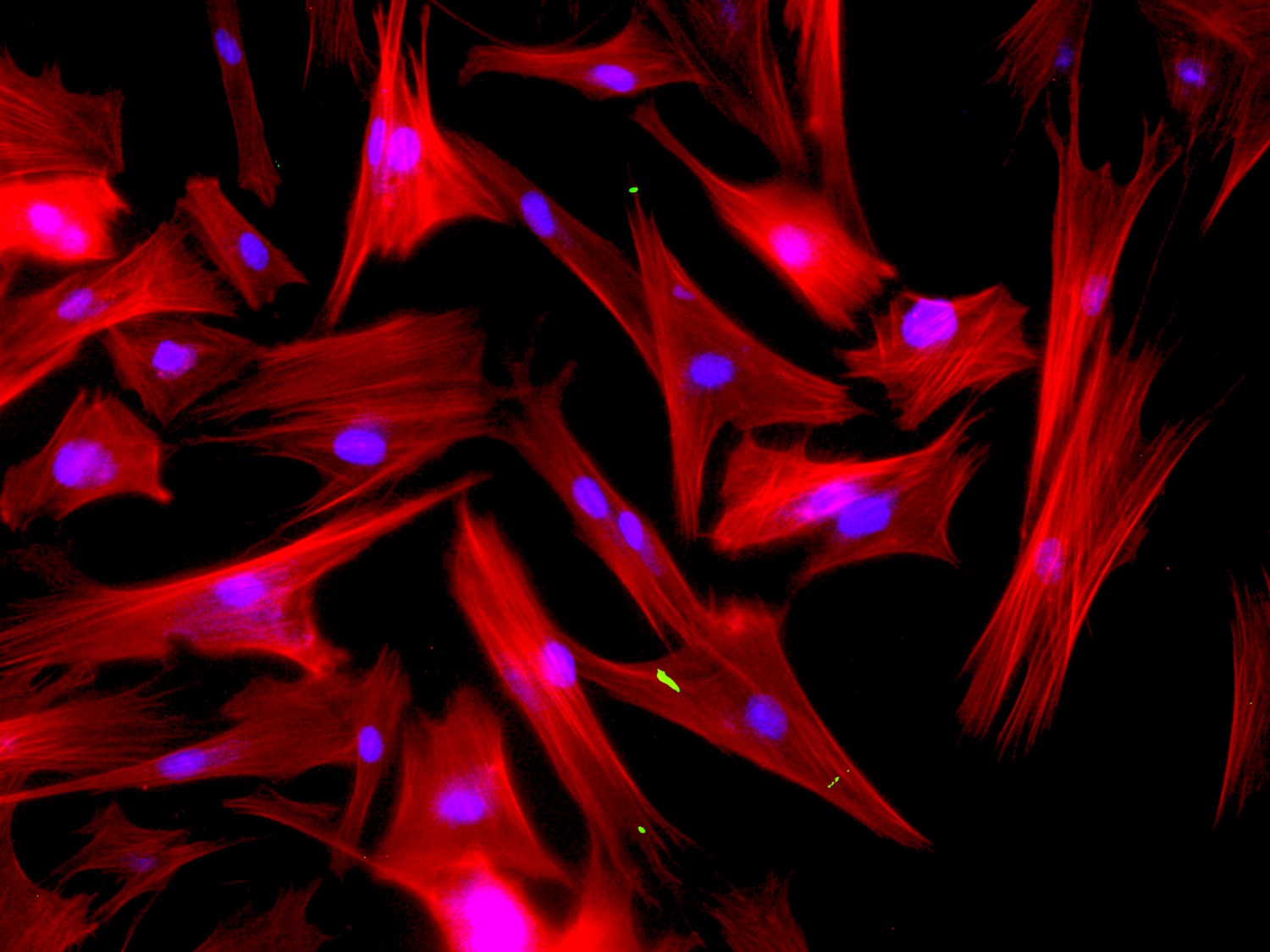
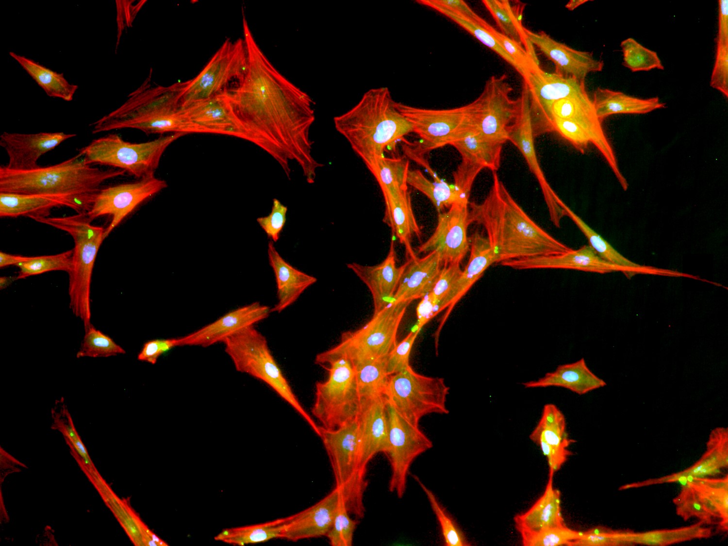
The core microscope imaging facility at PCMD was established as an interdepartmental facility to facilitate researcher to understand molecular mechanism working in the field of stem cells, microbiology, biotechnology, and cell biology of various diseases. Currently, the facility is equipped with state-of-the-art microscope and image analysis software that can be too expensive to purchase and maintain by any single institutions. The facility offers a wide range of conventional, light, florescence, atomic force microscopy with image analysis software. The core objective is to provide affordable and efficient access to advance imaging facility for researcher at our and any other research institutes. Beside microscopes, the facility also has tissue embedding machine, microtome, cryomicrotome and fume hood to process samples for histopathology, immunohistopathology and immunocytochemical analysis. Staff at the core imaging facility will provides assistance to use the installed microscopes.
The core microscope imaging facility at PCMD was established as an interdepartmental facility to facilitate researcher to understand molecular mechanism working in the field of stem cells, microbiology, biotechnology, and cell biology of various diseases. Currently, the facility is equipped with state-of-the-art microscope and image analysis software that can be too expensive to purchase and maintain by any single institutions. The facility offers a wide range of conventional, light, florescence, atomic force microscopy with image analysis software. The core objective is to provide affordable and efficient access to advance imaging facility for researcher at our and any other research institutes. Beside microscopes, the facility also has tissue embedding machine, microtome, cryomicrotome and fume hood to process samples for histopathology, immunohistopathology and immunocytochemical analysis. Staff at the core imaging facility will provides assistance to use the installed microscopes.
 |
 |
 |
 |
 |
 |
AVAILABLE MICROSCOPES
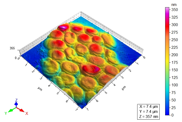
a:1:{s:7:"English";s:5219:"ATOMIC FORCE MICROSCOPE (AGILENT 5500):
It offers atomic resolution and is ideal for electrochemistry, polymers, and soft
material applications.
Features:
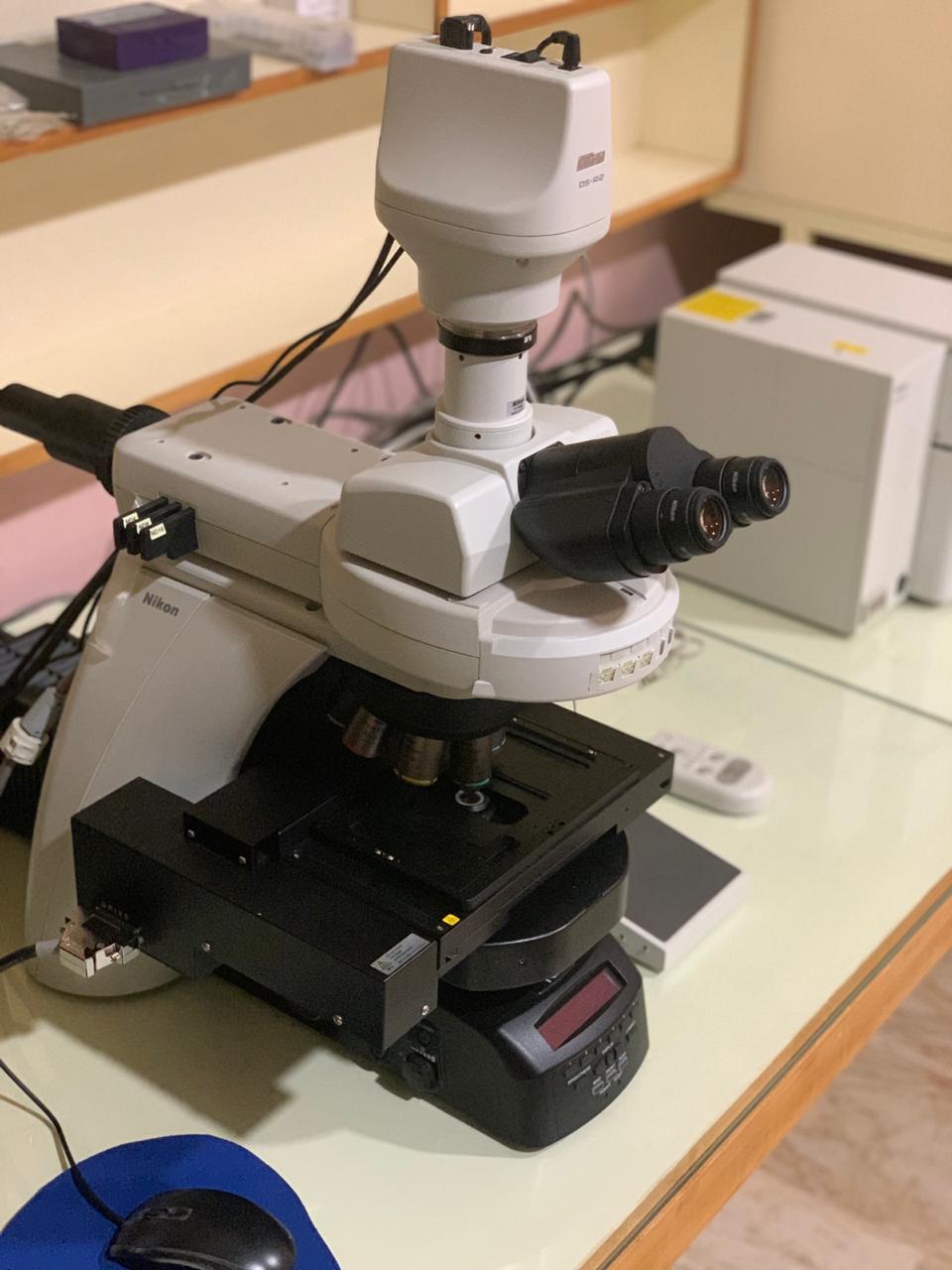
NIKON NI-E MICROSCOPE
Nikon Eclipse Ni-E microscope is one of the advanced motorized upright microscope to
attain demands for sophistication and automation of research.
Features:
NIKON ECLIPSE TI-2 MICROSCOPE
The Eclipse Ti-2 is inverted microscope which deliver high quality images with bright
LED illumination and time proven CFI optical system.
Features:
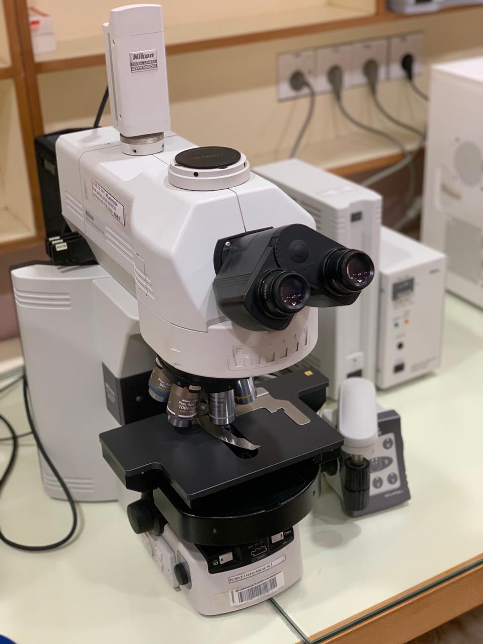
NIKON ECLIPSE 90i
Nikon Eclipse 90i is a motorized upright microscope imaging system with image
stitching function.
Features:
NIKON TE2000-E
Nikon Eclipse TE2000-E is a semi-motorized inverted microscope with NIS Elements software.
Features:
NIKON SMZ-800 STEREOMICROSCOPE
Stereo microscope allows 3D viewing of specimens.
Features:
";}
It offers atomic resolution and is ideal for electrochemistry, polymers, and soft
material applications.
Features:
- Patented top-down scanning
- Five modes: MAC, CSAFM, Contact, AAC, STM
- Temperature and environmental
- Control chamber
- A top-down optical axis through the scanner.
.jpg) |
NIKON NI-E MICROSCOPE
Nikon Eclipse Ni-E microscope is one of the advanced motorized upright microscope to
attain demands for sophistication and automation of research.
Features:
- Highly precise motorized focusing.
- Automatic alignment
- CFI plan apochromatic lambda series objective lens (4x, 10x, 20x, 40x and 100x).
- Phase Contrast lens (10x and 40x)
- Perform bright field, DIC, phase contrast and florescence imaging.
- A built-in “fly-eye” lens ensures uniform and bright illumination of the field of view,
- from edge-to-edge.
- Equipped with 16.25-megapixel high resolution Nikon DS-Ri2 camera.
 |
NIKON ECLIPSE TI-2 MICROSCOPE
The Eclipse Ti-2 is inverted microscope which deliver high quality images with bright
LED illumination and time proven CFI optical system.
Features:
- Its equipped with a quartz fly-eye lens.
- High-intensity LED lamp house
- Objectives (4x, 10x, 20x, 40x, 100x,).
- Equipped with phase contrast objectives (10x and 40x).
- Equipped with motorized external phase-contrast system and stage.
- Differential Interference and Advanced Modulation Contrast.
- NIS Element software (Advance research)
 |
NIKON ECLIPSE 90i
Nikon Eclipse 90i is a motorized upright microscope imaging system with image
stitching function.
Features:
- Adjustable eyepieces.
- Objectives (4x, 10x, 20x, 40x, 60x and 100x objectives)
- Possible contrast techniques are bright field, florescence and DIC.
- Digital camera (DXM-1200C).
- NIS Elements software and imaging PC
 |
NIKON TE2000-E
Nikon Eclipse TE2000-E is a semi-motorized inverted microscope with NIS Elements software.
Features:
- Adjustable Eyepieces and Switchable 1.5x Magnification Changer
- Objectives (4x, 10x, 20x, 40x and 60x).
- Possible contrast techniques are bright field, florescence and DIC.
- Available filters (DAPI, FITC and Texas Red).
- Digital camera (DS-5M and DS-2MBWc).
 |
NIKON SMZ-800 STEREOMICROSCOPE
Stereo microscope allows 3D viewing of specimens.
Features:
- The high-eye point eyepieces with diopter adjustment
 |
";}